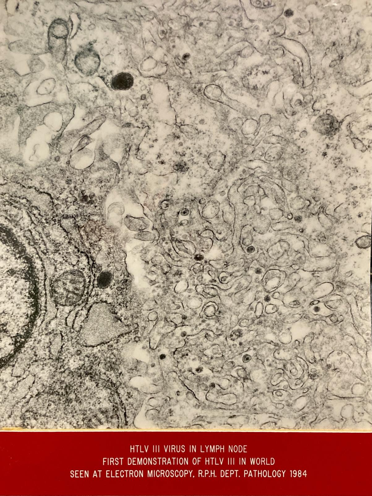
HIV VIRUS IN LYMPH NODE VIEWED THROUGH ELECTRON TRANSMISSION MICROSCOPE
1984Black & white photograph mounted on cardboard and displayed in a clear acrylic stand. The photograph depicts the HIV virus which was first visualised at RPH using the Electron Transmission Microscope.
There is a red label at the bottom of the stand which reads:
'HTLV 111 Virus in Lymph Node
First Demonstration of HTLV 111 in World
Seen at Electron Microscope, R.P.H. Dept. Pathology 1984'.
The Transmission Electron Microscope is still located in the RPH Pathology Department in Milligan House. It is no longer working.
Details
Details
RPH Pathology Department
Medical Research
AIDS (Acquired Immunodeficiency Syndrome) was first reported in the early 1980s and the disease quickly reached epidemic proportions.
Health workers at RPH became aware of the disorder within days of the early reports and quickly instituted diagnostic and therapeutic regimes to support those afflicted.
In addition to the Medical and Surgical Units that dealt with the epidemic, the Departments of Immunology, Microbiology, Medical Physics and Pathology focused on the peculiarities of the disease in their attempts to define its causation.
The Pathology Department’s Electron Microscopy Unit carefully scrutinised samples from affected patients. As well as clarifying how organs were affected during the course of the disease, they were able to detect, for the very first time in human tissues, the virus particles that came to be known as HIV (Human Immunodeficiency Virus).
Their ground-breaking findings were published in Lancet, one of the most prestigious medical journals.
Scientific, social and historic significance. First demonstration of HTLV III in the world.


